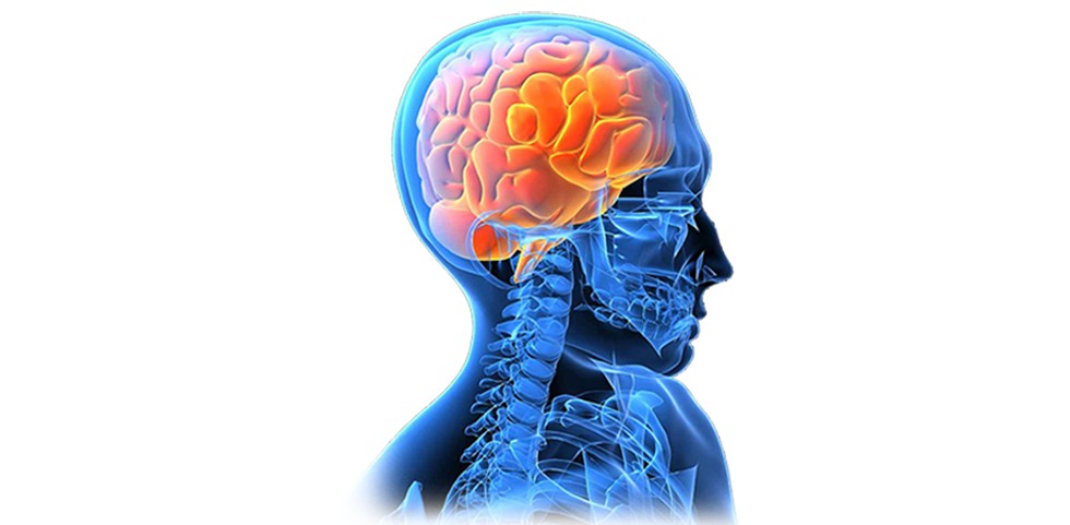
Brain Cancer
With a vision to offers comprehensive and state of the art Brain Cancer care, Dr. Vaibhav Choudahry provides the support & guidance patients and their families need with one-on-one consultations.
Sign & Symptoms:
People with a brain tumor may experience the following symptoms or signs. Sometimes, people with a brain tumor do not have any of these changes. Or, the cause of a symptom may be a different medical condition that is not a brain tumor.
Symptoms of a brain tumor can be general or specific. A general symptom is caused by the pressure of the tumor on the brain or spinal cord. Specific symptoms are caused when a specific part of the brain is not working well because of the tumor. For many people with a brain tumor, they were diagnosed when they went to the doctor after experiencing a problem, such as a headache or other changes.
- Headaches, which may be severe and worsen with activity or in the early morning
- Seizures. People may experience different types of seizures. Certain drugs can help prevent or control them. Motor seizures, also called convulsions, are sudden involuntary movements of a person’s muscles. The different types of seizures and what they look like are listed below:
1. Myoclonic
- Single or multiple muscle twitches, jerks, spasms
2. Tonic-Clonic (Grand Mal)
- Loss of consciousness and body tone, followed by twitching and relaxing muscles that are called contractions
- Loss of control of body functions, such as loss of bladder control
- May be a short 30-second period of no breathing and a person’s skin may turn a shade of blue, purple, gray, white, or green
- After this type of seizure, a person may be sleepy and experience a headache, confusion, weakness, numbness, and sore muscles.
3. Sensory
- Change in sensation, vision, smell, and/or hearing without losing consciousness
4. Complex partial
- May cause a loss of awareness or a partial or total loss of consciousness
- May be associated with repetitive, unintentional movements, such as twitching
- Personality or memory changes
- Nausea or vomiting
- Fatigue
- Drowsiness
- Sleep problems
- Memory problems
- Changes in ability to walk or perform daily activities
Symptoms that may be specific to the location of the tumor include:
- Pressure or headache near the tumor
- Loss of balance and difficulty with fine motor skills is linked with a tumor in the cerebellum.
- Changes in judgment, including loss of initiative, sluggishness, and muscle weakness or paralysis is associated with a tumor in the frontal lobe of the cerebrum.
- Partial or complete loss of vision is caused by a tumor in the occipital lobe or temporal lobe of the cerebrum.
- Changes in speech, hearing, memory, or emotional state, such as aggressiveness and problems understanding or retrieving words can develop from a tumor in the frontal and temporal lobe of the cerebrum.
- Altered perception of touch or pressure, arm or leg weakness on 1 side of the body, or confusion with left and right sides of the body are linked to a tumor in the frontal or parietal lobe of the cerebrum.
- Inability to look upward can be caused by a pineal gland tumor.
- Lactation, which is the secretion of breast milk, and altered menstrual periods in women, and growth in hands and feet in adults are linked with a pituitary tumor.
- Difficulty swallowing, facial weakness or numbness, or double vision is a symptom of a tumor in the brain stem.
- Vision changes, including loss of part of the vision or double vision can be from a tumor in the temporal lobe, occipital lobe, or brain stem.

Treatments Recommended :
Head and neck cancer specialists usually form a multidisciplinary team to care for each patient, and an evaluation should be done by each doctor before any treatment begins. This team often includes these specialists:
 Surgery
Surgery is the removal of the tumor and some surrounding healthy tissue during an operation. It is usually the first treatment used for a brain tumor. It is often the only treatment needed for a low-grade brain tumor. Removing the tumor can improve neurological symptoms, provide tissue for diagnosis and genetic analysis, help make other brain tumor treatments more effective, and, in many instances, improve the prognosis of a person with a brain tumor.
A neurosurgeon is a doctor who specializes in surgery on the brain and spinal column. Surgery to the brain requires the removal of part of the skull, a procedure called a craniotomy. After the surgeon removes the tumor, the patient’s own bone will be used to cover the opening in the skull.
There have been rapid advances in surgery for brain tumors, including the use of cortical mapping, enhanced imaging, and fluorescent dyes.
 Radiation therapy
Radiation therapy is the use of high-energy x-rays or other particles to destroy tumor cells. Doctors may use radiation therapy to slow or stop the growth of a brain tumor. It is typically given after surgery and possibly along with chemotherapy. A doctor who specializes in giving radiation therapy to treat a tumor is called a radiation oncologist. The most common type of radiation treatment is called external-beam radiation therapy, which is radiation given from a machine outside the body. When radiation treatment is given using implants, it is called internal radiation therapy or brachytherapy. A radiation therapy regimen, or schedule, usually consists of a specific number of treatments given over a set period of time.
External-beam radiation therapy can be directed at a brain tumor in the following ways:
- Conventional radiation therapy: The treatment location is determined based on anatomic landmarks and x-rays. In certain situations, such as whole brain radiation therapy for brain metastases, this technique is appropriate. For more precise targeting, different techniques are needed. The amount of radiation given depends on the tumor’s grade.
- 3-dimensional conformal radiation therapy (3D-CRT): Using images from CT and MRI scans (see Diagnosis), a 3-dimensional model of the tumor and healthy tissue surrounding the tumor is created on a computer. This model can be used to aim the radiation beams directly at the tumor, sparing the healthy tissue from high doses of radiation therapy.
- Intensity modulated radiation therapy (IMRT): IMRT is a type of 3D-CRT (see above) that can more directly target a tumor. It can deliver higher doses of radiation to the tumor while giving less to the surrounding healthy tissue. In IMRT, the radiation beams are broken up into smaller beams and the intensity of each of these smaller beams can be changed. This means that the more intense beams, or the beams giving more radiation, can be directed only at the tumor.
- Proton therapy: Proton therapy is a type of external-beam radiation therapy that uses protons rather than x-rays. At high energy, protons can destroy tumor cells. Proton beam therapy is typically used for tumors when less radiation is needed because of the location. This includes tumors that have grown into nearby bone, such as the base of skull, and those near the optic nerve.
- Stereotactic radiosurgery: Stereotactic radiosurgery is the use of a single, high dose of radiation given directly to the tumor and not healthy tissue. It works best for a tumor that is only in 1 area of the brain and certain noncancerous tumors. It can also be used when a person has more than 1 metastatic brain tumor. There are many different types of stereotactic radiosurgery equipment, including:
- A modified linear accelerator is a machine that creates high-energy radiation by using electricity to form a stream of fast-moving subatomic particles.
- A gamma knife is another form of radiation therapy that concentrates highly focused beams of gamma radiation on the tumor.
- A cyber knife is a robotic device used in radiation therapy to guide radiation to the tumor, particularly in the brain, head, and neck regions.
- Fractionated stereotactic radiation therapy: Radiation therapy is delivered with stereotactic precision but divided into small daily doses called fractions and given over several days or weeks, in contrast to the 1-day radiosurgery. This technique is used for tumors located close to sensitive structures, such as the optic nerves or brain stem.
With these different techniques, doctors are trying to be more precise and reduce radiation exposure to the surrounding healthy brain tissue. Depending on the size and location of the tumor, the radiation oncologist may choose any of the above radiation techniques. In certain situations, a combination of multiple techniques may work best.
Short-term side effects from radiation therapy may include fatigue, mild skin reactions, hair loss, upset stomach, and neurologic symptoms, such as memory problems. Most side effects go away soon after treatment is finished. Also, radiation therapy is usually not recommended for children younger than 5 because of the high risk of damage to their developing brains. Longer term side effects of radiation therapy depend on how much healthy tissue received radiation and include memory and hormonal problems and cognitive (thought process) changes, such as difficulty understanding and performing complex tasks.
 Chemotherapy
Chemotherapy is the use of drugs to destroy tumor cells, usually by keeping the tumor cells from growing, dividing, and making more cells.
A chemotherapy regimen, or schedule, usually consists of a specific number of cycles given over a set period of time. A patient may receive 1 drug at a time or a combination of different drugs given at the same time. The goal of chemotherapy can be to destroy tumor cells remaining after surgery, slow a tumor’s growth, or reduce symptoms.
As explained above, chemotherapy to treat a brain tumor is typically given after surgery and possibly with or after radiation therapy, particularly if the tumor has come back after initial treatment.
Some drugs are better at going through the blood-brain barrier. These are the drugs often used for a brain tumor.
- Gliadel wafers are a way to give the drug carmustine. These wafers are placed in the area where the tumor was removed during surgery.
- For people with glioblastoma and high-grade glioma, the latest standard of care is radiation therapy with daily low-dose temozolomide (Temodar). This is followed by monthly doses of temozolomide after radiation therapy for 6 months to 1 year.
- A combination of 3 drugs, lomustine (Gleostine), procarbazine (Matulane), and vincristine (Vincasar), have been used along with radiation therapy. This approach has helped lengthen the lives of patients with grade III oligodendroglioma with a 1p/19q co-deletion (see also, “Biogenetic markers” in the Grades and Prognostic Factors section) when given either before or right after radiation therapy. It has also been shown to lengthen lives of patients after radiation therapy for a low-grade tumor that could not be completely removed with surgery. Clinical trials on the use of chemotherapy to delay radiation therapy for patients with low-grade glioma are ongoing.
Patients are monitored with a brain MRI every 2 to 3 months while receiving active treatment. Then, the length of time between MRI scans increases depending on the tumor’s grade. Patients often have regular MRIs to monitor their health after treatment is finished and the tumor has not grown. If the tumor grows during treatment, other treatment options will be considered.
The side effects of chemotherapy depend on the individual and the dose used, but they can include fatigue, risk of infection, nausea and vomiting, hair loss, loss of appetite and diarrhea. These side effects usually go away after treatment is finished. Rarely, certain drugs may cause some hearing loss. Others may cause kidney damage. Patients may be given extra fluid by IV to protect their kidneys.
Learn more about the basics of chemotherapy.