Cervical Cancer

The development of cervical cancer is caused by cell proliferation observed in the cervix. Squamous cell carcinoma, the most prevalent type of cervical cancer, accounts for 70% of cases. Additionally, adenocarcinoma, a less frequent variety that begins higher in the cervix and is more difficult to identify, accounts for around 25% of cases.

Cervical screening is the only method to detect any abnormal growth in the cervix, as cervical cells in the precancerous stage are rarely known to present any symptoms.

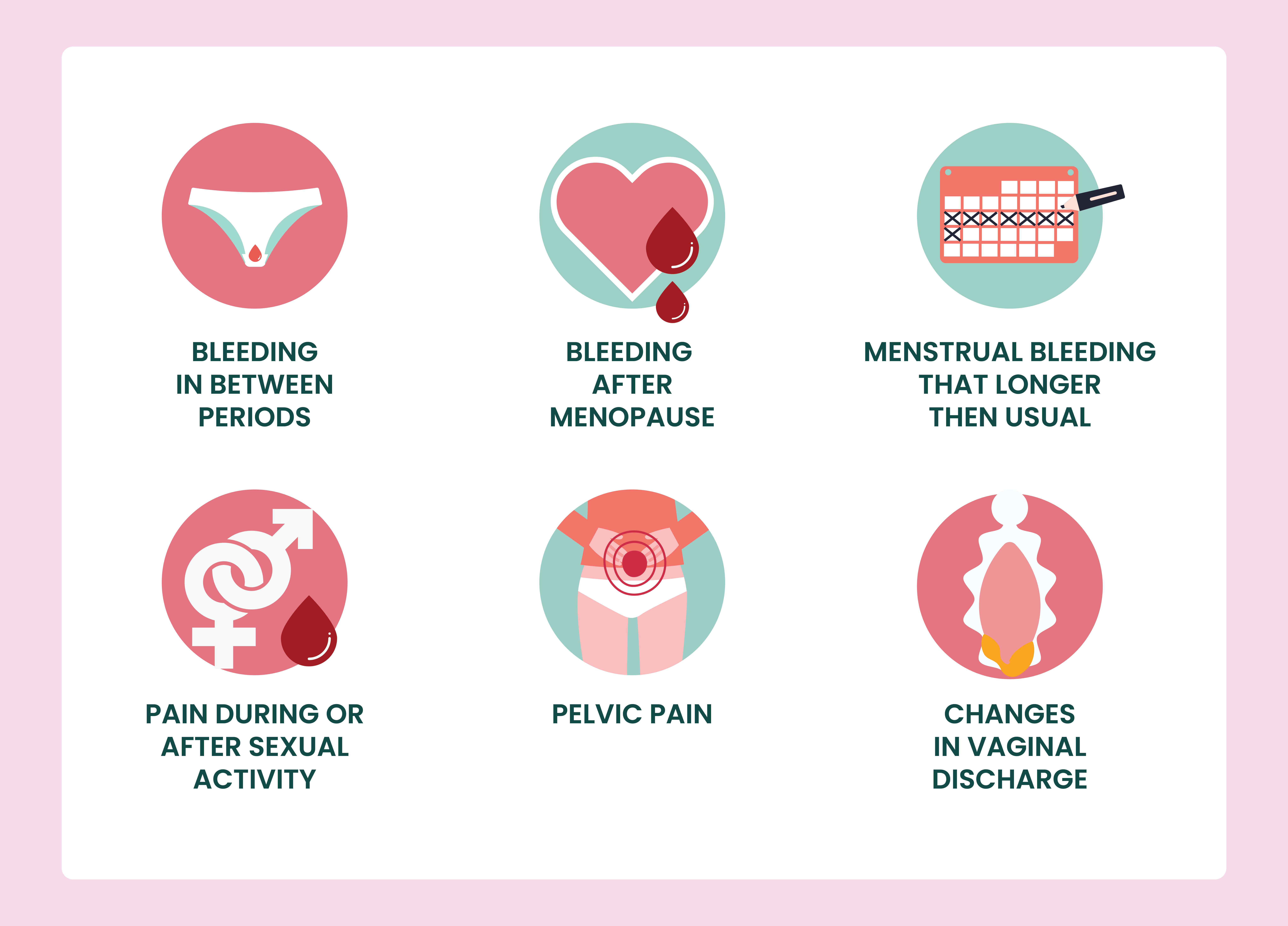
Among the most typical symptoms of the early cell alterations are:
  • Vaginal bleeding in between periods.
  • Vaginal bleeding after menopause
  • Menstrual bleeding that is greater or longer than normal
  • Pain during or after sexual activity
  • Pelvic pain
  • Changes in vaginal discharge, such as increased discharge or vaginal bleeding that has a strong or peculiar color or smell.

These similar symptoms can also be seen in other illnesses, however if any of the symptoms continue, consult Dr. Manasi Shah, the best cancer specialist in Ahmedabad.
If the findings of your screening indicate that you may have cervical cancer, more testing is done to confirm the diagnosis. These tests include:
  • Colposcopy combined with biopsy: Colposcopy is used to pinpoint the precise location and features of abnormal cells in the cervix. A tissue sample from the cervix's surface is collected for additional analysis if, during the process, any suspicious- looking area is noticed.
  • Cone biopsy: Cone biopsy,also known as large loop excision of the transformation zone (LLETZ), is the most popular technique for removing cervical tissue for testing and treating precancerous alterations in the cervix. Most of the time, a local anesthetic is used. When there are unusual glandular cells in the cervix or when early-stage cancer is suspected, a cone biopsy is performed.

If cervical cancer is discovered, it will be graded, going from stage 1, which indicates that abnormal cells are only present in the cervix's tissue, to stage 4, which indicates that the disease has progressed to the lung, liver, or bones in addition to the pelvis.

Choice of treatment varies according to illness stage. Treatment for non-bulky, early illness (less than 4 cm and node negative) involves surgery, followed occasionally by chemoradiation therapy.

A combination of chemotherapy and radiation therapy is utilized for locally advanced illness.

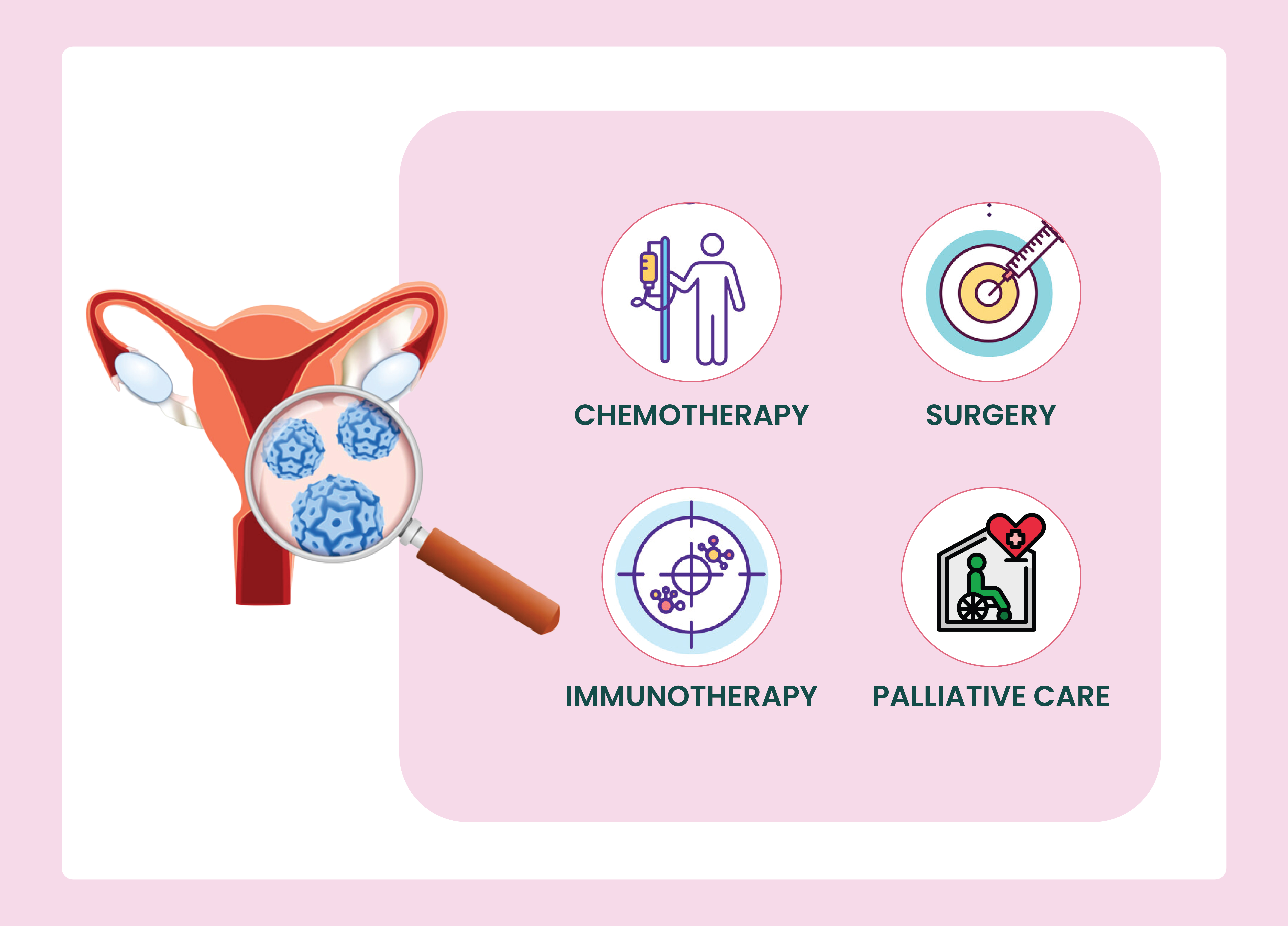
Treatment options for metastatic illness include chemotherapy, targeted therapy, immunotherapy and palliative care.